Objectives
You will be able to:
Understand the indications for using ultrasound guided access
Utilise various techniques for vascular access
Be able to accurately confirm IV placement
Understand the potential pitfalls and problems
Why do I need ultrasound?.....
The use of ultrasound has become THE standard of care in many line placement scenarios...the use of US has been shown over and over to improve precision, reduce numbers of attempts and reduce complications.
And one only needs to looks at the body habitus of our patients to realise that the depth and difficulty of IV line placement seems to be increasing as much as they are.
We will look at the use of ultrasound in both peripheral and central venous access.
USS GUIDED PERIPHERAL IV ACCESS
ULTRASOUND GUIDED PERIPHERAL IV (litfl)
USS GUIDED IV (5MIN SONO)
USS GUIDED PIV (EMDOCS)
USS GUIDED PIV (EM IN 5)
CENTRAL LINE PLACEMENT (5MIN SONO)
CENTRAL LINE CONFIRMATION (5MIN SONO)
Huge thanks to Dr Jennifer Martin and Dr Geoffrey Hayden for the kind use of their excellent lectures:
Peripheral Ultrasound Guided IV Access
Ultrasound Guided IV Access
Pearls and Pitfalls
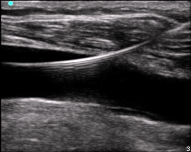
Failure to identify the needle in the tissue. Remember to look for the ring-down artifact to avoid this.
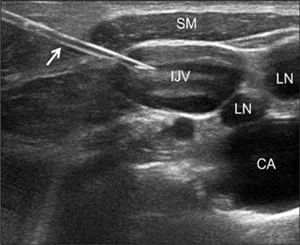
Failure to distinguish between vein and artery. Remember to look for the compressible vessel. Doppler flow can be used if necessary.
Angling the transducer towards the entry site of the needle on the skin may help visualize the needle earlier.
Avoid advancing the catheter if the needle tip is not visualized.
Placing the patient in a supine and Trendelenburg position will help facilitate central venous access.
Having the patient perform a Valsalva maneuver will help engorge the internal jugular vein.
Ideal positioning of neck should be midline or slightly lateral. Excessive head rotation may cause dangerous overlay of the internal jugular vein over the carotid artery.
Use caution when utilizing a long axis approach to central venous cannulation due to the inability to maintain visualization of the carotid artery at all times.
Estimate the length of the needle path and choose a catheter with the appropriate length. Failure to use sterile ultrasound gel for line placement. If not available you can substitute with a package of surgical lubricant.