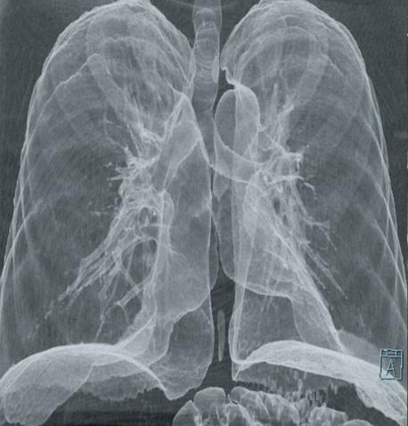
PoCUS LUNG ULTRASOUND
Lung ultrasound at the beside has the ability to diagnose most lung associated pathologies with a high degree of sensitivity and specificity. Performed systematically this often will out perform a CXR. Bedside lung ultrasound also allows you to attain helpful dynamic information about your patient over their clinical course.
Focused Acute Medicine Ultrasound (FAMUS) provides an excellent review of lung ultrasound
THORACIC ULTRASOUND THEORY (FAMUS)
Lung ultrasound relies heavily on the interpretation of artifacts, the artifact patterns, and pleural based findings.
AIMS
Understand the anatomy
Identify normal pleural sliding and lung ultrasound findings
Recognise and interpret ultrasound lung artifacts
A and B lines
Comet-tail and reverberation artifacts
Appreciate characteristic ultrasound findings of different pathologies
Pneumothorax
Interstitial syndrome - ‘Wet lungs’
Alvelolar syndrome - Consolidation
Pleural effusions
LUNG ULTRASOUND INDICATIONS
Lung ultrasound has broad application in differentiating causes of respiratory distress and dyspnea. It can also be integrated into shock and volume assessments:
Pneumothorax
Causes of interstitial syndrome (‘Wet lungs’) e.g cardiogenic pulmonary edema
Pneumonia
Pleural effusions
Part of a shock and volume assessment
LIMITATIONS
Extensive surgical emphysema will impair views
Large body habitus
Patient mobility/positioning limitations may be an issue
LUNG ULTRASOUND PROTOCOLS
A number of approaches to ED based lung ultrasound have been developed.
The most recognised is the BLUE (Bedside Lung Ultrasound in Emergency) Protocol (Lichtenstein 2008).
This utilises three imaging points on each sides of the chest that helps to define specific ultrasound profiles in conditions causing dyspnea
The BLUE protocol can be reasonably confusing approach if not familiar
BLUE PROTOCOL IN DETAIL
In general a more simplified approach can be taken utilising a 6-point lung imaging technique, looking for:
Presence or absence of lung sliding (+ M-Mode)
Is there a pneumothorax ( You may find a lung point)
Other causes of loss of lung sliding
B-lines
> 3 per field of view abnormal
Localised vs diffuse
Is there an interstitial syndrome
Looking for signs of consolidation
Looking for effusions
This can be integrated with an IVC and gross cardiac function assessment to help determine cause of any identified interstitial syndrome i.e. Diffuse B-lines seen in cardiogenic pulmonary edema
ULTRASOUND APPEARANCE OF LUNG
In normal air filled lung, ultrasound can only interrogate the pleural surface. The ultrasound waves are near completely reflected/attenuated at the normal pleural/air interface:
In the normal lung everything below the pleural line is grey-scale artifact filled in by the ultrasound machine
These artifacts are useful in lung ultrasound
Normal lung parenchyma is not seen in ultrasound
Interstitial fluid accumulation results in potentially diagnostic artifactual B-lines
Pathologic change can result in visualisation of areas of lung parenchyma by ultrasound
Fluid displaces air in alveoli/airways
Collapse and consolidation of lung occurs
Pleural effusions are seen as anechoiec (black) accumulations
A number of described ultrasound signs can be seen with particular lung pathologies.
SURFACE ANATOMY
Source: POCUS 101
Source: POCUS 101
LUNG ULTRASOUND PROBES AND PRESETS
Pleural and superficial assessment:
High frequency linear probe is best for this, set at a depth of 4-6 cm, with the focal point at the pleura level
Detection of pneumothorax - Lung sliding and M-mode
Pleural changes and pleural based pneumonic changes
General lung imaging:
Curvilinear probe is probably the best option set at a depth of around 15cm
Can see pleural sliding (Not as good as the linear probe)
Has wide field of view
Has good resolution at depth
Phased probe is also used widely owing to its small footprint allowing imaging within small acoustic windows of the chest
Less detail of superficial lung and pleura
Poorer spatial resolution at depth compared to the curvilinear probe
Presets:
A lung preset should be used initially in your exam
Turns off tissue harmonic imaging (THI) and multi-beam (MB)/compound imaging which accentuates important diagnostic lung artifacts (e.g. B-lines)
An Abdo preset can be used for more detailed imaging of pleural effusions, and areas of parenchymal abnormalities such as lung consolidations
THI and MB are activated improving image resolution and reduces artifact
SIX-point lung ultrasound
LUNG ULTRASOUND MADE EASY (POCUS 101)
An initial six-point (3 per side) approach is probably sufficient for initial beside lung ultrasound
A more extensive examination, including the posterior chest, can be undertaken if looking for more localised findings such as pneumonia
There are various techniques for a more comprehensive exam
COMPREHENSIVE LUNG EXAM (Life in The Fastlane)
Patient positioning:
Generally the patient will be supine or reclined in the bed for a 6-point exam
In the patient with more marked respiratory distress they may be upright
More detailed/extensive imaging would require the patient to be seated upright with the back away from the bed
PROBE POINTS
3-points imaged on each of the right and left sides:
Probe is in the longitudinal plane with probe marker to the head
R1/L1: mid-clavicular line at the 2-3rd intercostal space
Correlates to the R and L upper lobes of the lung
R2/L2: mid-axillary line at the 6-7th intercostal spaces (lateral to male nipple level)
Correlates to the R middle lobe and lingula lobe on the left
R3/L3 : posterior-axillary line at the 10-11th intercostal space (Xiphoid level)
Correlates to the R and L lower lobes
R3/L3 known as the PLAPS points (posterior and/or lateral alveolar and/or pleural syndrome)
Most common place to find lung consolidation and pleural effusions
Source: POCUS 101
At each point identify ribs, pleural line and adjust depth appropriately
Identify the ‘BAT WING SIGN’ at each point
Identify lung sliding at each point
Source: Westersono (Modified)
ANTERIOR CHEST R1/L1
2-3rd intercostal space in the mid-clavicular line
Most anterior/apical part of the chest (in the near supine patient)
You can use the linear probe initially to look for pleural sliding and then change to curvilinear probe for examination at depth
Location for looking for a pneumothorax and also B-lines
Need to identify normal lung sliding
Use M-mode to identify the normal ‘Seashore’ sign pattern
Note other normal lung artifacts
A lines (reverberation artifact) repetitions of the pleural line at depth
Present in normal and abnormal lungs
Not abolished by a pneumothorax
Most prominent when the probe is perpendicular to pleura
Short pleural based comet-tail artifacts (seen in lung sliding video above)
LATERAL CHEST R2/L2
6-7th intercostal space in the mid-axillary line
Locate ‘BAT WING’ sign , pleural sliding , and A lines
Identify any abnormalities
POSTERIOR CHEST R3/L3 PLAPS POINT
This is the location of interest where you are most likely to see effusions or consolidation
Place the probe as near to the bed as possible, in line with the xiphoid using the liver (R3) and spleen (L3) as sonic windows.
This equates to a similar position as the RUQ and LUQ eFAST views
You should note the typical normal findings
Mirror imaging artifact of liver or spleen above the diaphragm
The image of the spine stopping at the diaphragm level as it heads cephalad
The ‘CURTAIN SIGN’ of normal aerated lung sliding down with inspiration in to the frame
Scan slightly cephalad interrogating along the posterior axillary line
LUNG ULTRASOUND FINDINGS
COMMON LUNG ULTRASOUND FINDINGS
PNEUMOTHORAX
Key to identifying a pneumothorax is to scan the anterior most aspect of the chest, in the supine patient this correlates to the R1/L1 position in the 6-point examination.
Remember the findings are location specific to the probe position, but if the probe is at the most anterior portion/or apical portion of the chest a significant pneumothorax should be detected.
Pneumothorax RULE OUT (at the imaged location) - normal lung sliding (NPV 99%)
Use M-mode to help confirm your findings
‘SEASHORE SIGN’ is normal (see above)
‘STRATOSPHERE SIGN’ is consistent with a pneumothorax (see below)
Source: (Modified)
Absence of lung sliding in the right clinical context is suggestive of a pneumothorax
Other causes of apparent absence of lung sliding:
Apnoea/respiratory arrest
Poorly ventilated patients
Severe hyperinflation - asthma/COPD
Adhesions/consolidation
Pleurodesis
Blebs or bullae
The presence of B-lines or a lung pulse exclude a pneumothorax at that point
A lung pulse may be seen in conditions where there is apparent loss of normal pleural sliding but without a pneumothorax
A lung pulsation seen at the pleura is the transmitted pulsation/movement from the heart
Usually seen when there is absent/poor ventilation, apnoea, or severe hyper- inflation
Finding a lung-point (PPV 100%) is a ‘RULE IN’ for a pneumothorax
A lung-point is found by scanning progressively more lateral and caudad in the chest
Location of the lung point confirms the diagnosis, and helps to quantify the size.
It is important not to mistakenly interpret a lung point from a pseudo-lung point (video below)
A pseudo-lung point is a junction between the lung and mediastinum or diaphragm
Often seen on the left side of the chest where lung meets pericardium
PSEUDO-LUNG POINT VS LUNG POINT (Dr James Rippey)
B-LINES AND INTERSTITIAL SYNDROME
B- lines are a vertical reverberation artifact that results from changes in the interstitial lung tissue
Caused be increased fluid and/or thickening of the interstitial space of the subpleural and interlobular septa
Variety of conditions can generate B-lines which may be focal or diffuse in nature
Source: Radiology Key
b-line Ultrasound Definition
Vertical reverberation artifact
Originate from the pleura
Extend to beyond the maximal depth of view
Hyperechoiec
Move with pleural sliding
POCUS GEEK - B Lines and Hepitisation
causes of b-lines
A few ‘normal’ B lines can be commonly found especially in the older age groups
Typically in dependent locations (Lung bases)
< 3 per field of view can be normal
Usually the B-lines will not be diffuse in this setting
Abnormal B-Lines
3 or more B-lines per field of view (intercostal space) is abnormal
These may be focal or diffuse depending on the pathology
Diffuse B-lines in 3 or more zones on both sides of the chest is consistent with a diffuse alveolar interstitial syndrome ‘WET LUNGS’ such as pulmonary oedema or ARDS
B-lines may be seen in a number of chronic conditions causing interstitial thickening
Pleural fibrosis from any cause
Interstitial lung disease/fibrosis
B-lines will be seen in a pattern of distribution related the areas of the lung affected by the underlying condition
Typically more localised to certain zones
Cardiogenic Pulmonary Oedema
Typically bilateral B-lines
Associated with cardiac dysfunction on bedside echo
Mild overload states B-lines are found in dependent locations
In more acute moderate-severe cases they will become diffuse
B-lines may become confluent with each other with severe ‘WET LUNG’
ARDS
Any cause of ARDS will produce B-lines
Generally diffuse and bilateral
Pneumonia
May be focal or diffuse/bilateral or unilateral depending on pattern of pneumonia
Typically focal associated with subpleural or lobar consolidations
Trauma
Pulmonary contusion will lead to B-lines which are typically localised to the area of injury
May be associated with other findings such as lung consolidation from haemorrhage, pleural effusion from haemothorax, or pneumothorax.
consolidation and alveolar syndrome
Normal aerated lung parenchyma is not seen on ultrasound. Consolidated lung becomes apparent on ultrasound, when fluid accumulation and lung collapse results in displacement of air from the tissue.
Consolidation of lung may occur for a variety of reasons:
Pneumonia
Extrinsic mass effect causing collapse e.g. large pleural effusion
Intrinsic obstruction causing collapse e.g. bronchial mass, mucous plugging etc.
Significant atelectasis
CONSOLIDATION IN PNEUMONIA
The ultrasound changes seen in pneumonia are a spectrum of progressive changes depending on the severity and the extent of the process:
WesternSono- Alveolar Syndrome
ULTRASOUND FINDINGS IN PNEUMONIA
There are a variety of ultrasound findings in pneumonia:
B-lines may be focal or diffuse, unilateral or bilateral
Pleural thickening
Sub-pleural consolidations
Hypoechoeic sub-pleural fluid/pus filled foci surrounded by hyperechoeic border
Source: BMC Paediatrics
Shred sign
Hyperechoeic demarcation between normal aerated lung and consolidated lung
Source: Dr Mark Tessaro
Air-Bronchograms
Air is trapped in consolidated or collapsed lung tissue
These may be static or dynamic in nature
Static air trapped may be found in atelectasis or consolidation
Dynamic air is still in communication and is usually found in consolidated lung
Shred sign and Static Air Bronchogram
Source- ACEP
Dynamic Air Bronchogram
Source- ACEP
Dense consolidation
Hepatisation (‘Liver like’) of the lung
See video below dense consolidated lung above (to the left) of the diaphragm, with the liver below (to the right). Bright echogenic shred sign between consolidated lung and aerated lung
Hepatisation
Source- ACEP
PLEURAL EFFUSIONS
Pleural Effusion Ultrasound
Source- Soundbytes Cases
Pleural effusions are typically identified most easily at the PLAPS (R3) point in a supine patient.
There are a number of signs that can help identify a pleural effusion and may also help determine the nature of the effusion within the clinical context i.e. transudate vs exudate.
Pleural fluid will typically appear relatively anechoeic in nature, found between the confines of the parietal pleura on the chest wall and the visceral pleura overlying the lungs.
The nature of the effusion will determine the relative appearance:
Transudatve effusion and acute haemothorax will appear black/anechoeic
Exudative effusions will tend to have a variable degree of echogenicity or ‘debri’
Loculated effusions which tend to be caused by inflammatory processes, and usually will be exudative in nature
Haemothorax can appear as an acute black/anechoeic fluid, mixed echogenicity, or almost tissue like if the blood is substantially clotted.
Haemothrorax
Source- ACEP
Loculated Effusion
Source- ACEP
pleural effusion size
An approximate pleural effusion volume can be measured using technique described by Balik et al. at the PLAPS point:
Choose measurement A or B which ever is the greatest
Small effusions measuring < 10mm less accurate
Loculated effusions will not be accurately measured
Pleural volume (mL) = (measured distance in mm) x 20.
POCUS PLEURAL EFFUSION SIGNS
Spine sign
In pleural effusion or lung consolidation, ultrasound waves can travel through the thoracic cavity above the diaphragm allowing the spine to be seen
Spine Sign
Plankton sign
Echogenic debri within the pleural effusion may be seen in exudative effusion or haemothorax
Plankton Sign
Source- POCUS 101
Jellyfish Sign
At the PLAPS point this is the appearance of consolidated lung floating in the pleural effusion
Jellyfish Sign
Quad Sign
Boundaries of a pleural effusion in a supine patient tends to make a ‘Quadrilateral Shape’ (see below) with the boundaries being the rib shadow either side, the superficial boundary being the parietal pleura, and the deep boundary being the visceral pleura over the lung.
Often B-lines will be visible deep to the effusion
Quad Sign
Sinusoid Sign
This is gained using M-mode through an apparent pleural effusion
This can help confirm the presence of a pleural effusion, which would be represented by a sinusoidal pattern of the lung moving towards the line of sight/probe during inspiration (see below).
Sinusoid Sign
Sinusoid Sign
Source- ERC


